What is Cornea Edema (Bullous Keratopathy)? Causes, Symptoms, and Advanced Treatment in Japan
- Dec 10, 2025
- 4 min read

I. Introduction: What is Corneal Edema? The Loss of Transparency
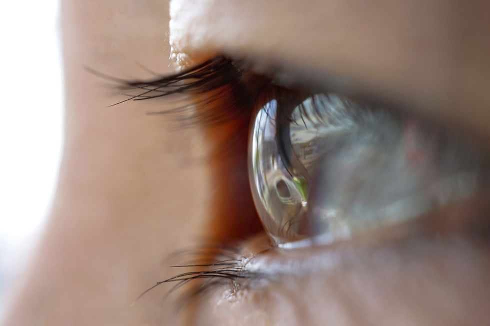
The clarity of the cornea, the eye's front window, is essential for sharp vision. Corneal edema is a condition where excess fluid accumulates within the corneal tissue, causing it to swell and lose its transparency. The severe, chronic manifestation of this condition is known as Bullous Keratopathy, which can lead to significant pain and blindness.
Understanding the underlying mechanism—the failure of the corneal endothelial cells—is crucial for effective treatment. This article provides a comprehensive overview of corneal edema and highlights how Japan's leadership in regenerative medicine is offering revolutionary solutions that move beyond traditional corneal transplantation.
II. The Mechanism & Causes: What causes cornea edema? The Endothelial Pump Failure
To understand what causes cornea edema, one must first understand the function of the corneal endothelium.
1. Endothelial Cell Function (The Pump):
The endothelium is a single, non-regenerative layer of cells lining the inner surface of the cornea. Its primary role is to act as a metabolic "pump," constantly drawing water out of the corneal stroma (the main structural layer). This pumping action maintains the cornea's optimal state of dehydration, which is necessary for its transparency.
2. Primary Mechanism (what causes cornea edema):
Corneal edema occurs when the number of viable endothelial cells falls below a critical threshold (typically 500 cells/mm^2), resulting in the pump's failure. When fluid cannot be expelled rapidly enough, it builds up, causing swelling and clouding.
3. Common Etiologies:
Fuchs' Endothelial Dystrophy (FED): A genetic, age-related condition where endothelial cells prematurely die or become dysfunctional.
Aging: Natural, progressive loss of endothelial cells over time.
Surgical Trauma: Damage to the endothelium during intraocular procedures (discussed in detail below).
III. Associated Symptoms
Early diagnosis is vital. Recognizing corneal edema symptoms can prevent the condition from progressing to severe Bullous Keratopathy.
1. Core Visual Symptoms:
Blurred or Hazy Vision: Often described as looking through steam or a fog.
Glare and Halos: Increased scattering of light, causing a shimmering effect or rings around light sources.
Fluctuating Vision: Vision may be worse in the morning (due to reduced evaporation while the eyes are closed overnight) and slowly improve throughout the day.
2. Physical Symptoms:
Ocular Pain and Discomfort: A constant foreign body sensation.
Photophobia: Sensitivity to light.
3. Progression to Bullous Keratopathy:
As the edema worsens, fluid accumulates under the surface layer (epithelium), forming fluid-filled blisters (bullae). When these bullae rupture, they expose nerve endings, causing intense, debilitating pain—the characteristic symptom of severe Bullous Keratopathy.
IV. Surgical Inducement
A significant cause of endothelial cell loss is iatrogenic damage (caused by medical intervention), particularly during intraocular surgery.
1. Cataract Surgery (corneal edema after cataract surgery):
Even highly successful cataract surgery carries a risk of endothelial cell loss. Damage is typically caused by:
Phaco Energy: The ultrasonic energy used to break up the cataract (Cumulative Dissipated Energy, CDE).
Instrument Contact: Direct mechanical contact with the fragile endothelial layer.
Inflammation: Post-operative inflammation.
Japanese surgeons minimize the risk of corneal edema after cataract surgery by utilizing advanced techniques and highly refined fluidics management to ensure the lowest possible CDE.
2. Glaucoma Surgery (corneal edema after glaucoma surgery):
Certain procedures to lower intraocular pressure (IOP) also pose a risk. Corneal edema after glaucoma surgery can occur, particularly when drainage devices or surgical manipulations are performed close to the angle structures near the peripheral cornea, where the endothelium is susceptible to damage.
V. Treatment Landscape: Corneal Edema Treatments
Treatment for corneal edema is tiered, ranging from conservative management to definitive surgical intervention.
1. Conservative and Medical Management:
Initial corneal edema treatments aim to temporarily alleviate symptoms:
Hypertonic Agents (corneal edema eye drops): Prescription corneal edema eye drops (e.g., 5% sodium chloride/hypertonic saline) work by drawing excess water out of the cornea through osmotic pressure. This offers symptomatic relief, particularly in the mornings, but does not address the root cause.
Soft Contact Lenses: Used as a bandage to reduce pain caused by ruptured bullae.
2. Surgical Management:
Until recently, the only definitive solution was replacement of the damaged cell layer via corneal edema transplantation:
Traditional Penetrating Keratoplasty (PKP): Full-thickness corneal replacement.
Endothelial Keratoplasty (DMEK/DSAEK): Partial thickness transplantation where only the diseased endothelium is replaced. While safer than PKP, these procedures still rely on donor tissue and carry a lifelong risk of immune rejection.
VI. The Japanese Innovation: Cell and Regenerative Therapy
Japan is leading the global effort to replace transplantation with non-donor-dependent, minimally invasive cell therapies.
1. Cell Therapy:
The frontier of cornea edema stem cell research has culminated in treatments like Neltependocel (Cultivated Endothelial Cell Injection). This procedure involves:
Cultivation: Harvesting healthy cells from a single donor cornea and cultivating them into sufficient numbers to treat multiple patients.
Injection: Injecting the cultivated cells into the patient's anterior chamber. The cells then adhere to the inner cornea, regenerating the pump function. This low-invasive technique significantly reduces the reliance on traditional donor supply.
2. The Role of iPS Cells:
Beyond using cultivated donor cells, Japanese research is pioneering the use of Induced Pluripotent Stem (iPS) Cells to generate endothelial cells in unlimited quantities. This research promises the ultimate solution: cell therapy that is entirely independent of donor availability and carries the lowest possible risk of rejection.
VII. Conclusion: Achieving Cornea Clarity: Hope in Advanced Regeneration
Corneal edema is a serious condition stemming from the failure of the corneal endothelial pump, with severe stages classified as Bullous Keratopathy.
The future of treating this condition lies in regeneration. Japanese precision in diagnosis and its unwavering commitment to cell therapy (including Neltependocel and iPS cell research) offer the most advanced, least invasive, and sustainable solutions for restoring ocular clarity and eliminating the pain associated with severe Bullous Keratopathy.
This article was reviewed by
Dr. Daiki Sakai, MD