What Are the 3 Types of Cataract Surgery? A Comprehensive Guide to Phacoemulsification, ECCE, and FLACS in Modern Ophthalmology
- Nov 11, 2025
- 5 min read

I. Introduction: What are the 3 types of Cataract Surgery?
Cataract surgery is one of the most frequently performed and successful procedures globally. Yet, the method has evolved dramatically, moving from highly invasive techniques to ultra-precise, micro-incisional approaches. To understand the options available for vision correction, it is essential to know the primary techniques: Phacoemulsification, Extracapsular Cataract Extraction (ECCE), and Femtosecond Laser-Assisted Cataract Surgery (FLACS).
This article provides a comprehensive guide to what are the 3 types of cataract surgery, detailing their mechanisms, applications, and why Japanese ophthalmologists meticulously select the appropriate method to ensure maximum patient safety and optimal visual outcomes.
II. Type 1: Phacoemulsification (The Modern Gold Standard)
Phacoemulsification, often simply called "Phaco," is the technique that revolutionized cataract surgery and remains the global gold standard for most cases today.
1. Mechanism:
The procedure involves creating a tiny, self-sealing micro-incision (typically 2.0mm to 3.0mm) in the cornea. A specialized probe is inserted through this opening. This probe uses high-frequency ultrasonic vibrations to emulsify (break up) the hard cataractous lens nucleus into small fragments. These fragments are then simultaneously suctioned (aspirated) out of the eye. Once the lens material is removed, the Intraocular Lens (IOL) is injected, usually in a folded state, through the same small incision and unfolds inside the capsular bag.
2. Key Advantage:
The main advantage of Phacoemulsification is the small incision size. This leads to:
Rapid Healing: The small, clear corneal incision generally heals quickly without the need for sutures.
Reduced Induced Astigmatism: Minimal distortion to the corneal structure compared to older techniques.
Quick Visual Recovery: Patients typically notice improved vision within days.
3. Application:
Phacoemulsification is effective for early-to-moderate grades of cataracts and is the technique of choice in most industrialized nations, including Japan.
III. Type 2: ECCE (The Traditional Method)
ECCE is the predecessor to Phacoemulsification. While it has been largely superseded by modern techniques, it still maintains a role in specific, limited clinical scenarios.
1. Mechanism:
Unlike Phaco, ECCE requires a larger incision (up to 10mm) in the cornea or sclera. The surgeon manually removes the entire intact lens nucleus from the eye in one piece, leaving the lens capsule (the bag holding the IOL) intact. The larger incision must be carefully closed with sutures afterward.
2. Application:
ECCE is now reserved primarily for:
Mature or Hyper-Mature Cataracts: Cases where the lens nucleus is extremely hard (Type IV in the Emily-Little classification) and cannot be safely broken down by ultrasound, or where the eye is fragile.
Traumatic Cataracts: Cases with zonular dialysis (weakened supporting fibers) where the small incision Phaco technique may be too strenuous.
Limited Resource Settings: Where Phaco equipment is unavailable.
3. Disadvantage:
The significant drawback is the large incision, which results in:
Slower Recovery: Sutures are required, increasing the risk of infection.
High Induced Astigmatism: The incision often causes significant changes in the corneal shape.
Longer Post-Operative Rehabilitation: Visual recovery is slower than Phaco or FLACS.
IV. Type 3: FLACS (The Innovative Future)
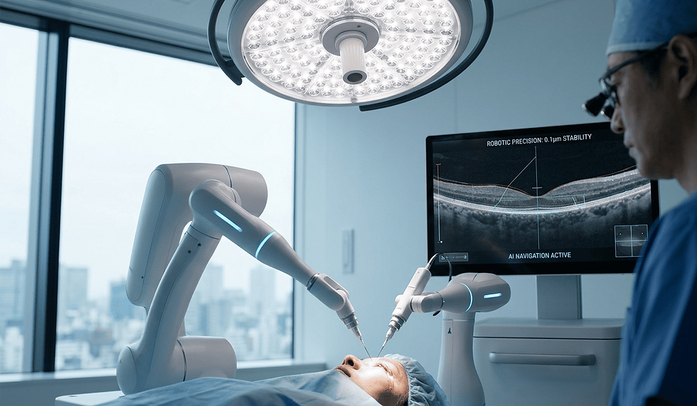
FLACS represents the most significant technological leap in cataract surgery in the last two decades. It integrates a Femtosecond laser to automate and refine the most critical steps of the procedure.
1. Mechanism:
FLACS does not replace Phacoemulsification entirely; rather, it performs the initial steps with unparalleled precision before the ultrasound probe is used. The laser executes three key maneuvers:
Corneal Incision: Creating highly precise, self-sealing incisions with consistent dimensions.
Capsulotomy: Creating a perfectly circular, centered opening in the lens capsule (capsulorhexis). This circularity is crucial for IOL stability.
Lens Fragmentation: Softening and fragmenting the hard lens nucleus before the Phaco probe is inserted, significantly reducing the amount of ultrasonic energy required.
2. Key Advantage:
The advantages stem from the laser's micron-level accuracy and high reproducibility:
Optimal IOL Positioning (Centration): The perfectly circular capsulotomy ensures the IOL rests precisely in the center of the visual axis, which is vital for the performance of Premium IOLs (multifocal and extended depth of focus lenses).
Minimized Stress: Pre-fragmentation dramatically reduces the necessary ultrasound energy, leading to lower corneal stress and a potentially quicker recovery of the cornea.
3. Application:
FLACS is recommended in advanced Japanese clinics for:
Patients receiving Premium IOLs who demand the highest refractive accuracy.
Patients with high astigmatism (where laser precision is used for Limbal Relaxing Incisions).
Patients with endothelial compromise or high-grade cataracts to minimize corneal risk.
V. Comparative Analysis
The evolution from ECCE to FLACS tracks a clear movement towards smaller incisions, lower energy, and higher precision.
Feature | ECCE (Extracapsular) | Phacoemulsification | FLACS (Femtosecond Laser-Assisted) |
Incision Size | Large (up to 10mm) | Micro-Incision (2.0-3.0mm) | Micro-Incision (Laser-made) |
Sutures Required? | Yes | Rarely | No (Self-sealing) |
Lens Removal | Manual removal of intact nucleus | Ultrasound fragmentation and aspiration | Laser pre-fragmentation, then aspiration (with reduced ultrasound) |
Precision (Capsulotomy) | Manual (Variable) | Manual (Variable) | Automated, near-perfect circle |
Energy Use | Zero ultrasound, but high mechanical stress | Moderate-to-High Ultrasound Energy | Lowest Ultrasound Energy |
Visual Recovery | Slow (Weeks to Months) | Fast (Days to a Week) | Fastest (Days) |
VI. The Japanese Expert Selection
Japanese ophthalmology is characterized by its rigorous safety protocols and keen adoption of technology, influencing the selection among the 3 types of cataract surgery.
1. Preference for FLACS/Phaco:
Due to the paramount focus on patient safety and minimizing post-operative astigmatism, ECCE is rarely performed in Japan, reserved only for extreme cases. The vast majority of procedures are either Phacoemulsification or, increasingly, FLACS.
2. FLACS for Premium IOLs:
Japan has a high rate of adoption for Premium IOLs, driven by patient demand for freedom from glasses after surgery. The superior IOL centration and stability provided by the laser's perfect capsulotomy make FLACS the preferred choice for these advanced lenses, maximizing their performance and the Quality of Vision (QOV) delivered to the patient.
3. Risk Mitigation:
Japanese specialists emphasize the reduction of ultrasonic energy, especially for higher grades of cataracts. By utilizing FLACS for pre-fragmentation, surgeons achieve the highest level of nuclear removal efficiency while maintaining the health of the delicate corneal endothelial cells—a commitment to long-term eye health.
VII. Conclusion: Precision Defined by Choice: The Best Approach for Your Cataract
The question of what are the 3 types of cataract surgery is answered by understanding surgical evolution: ECCE serves as a historical and fallback method, Phacoemulsification is the reliable standard, and FLACS is the cutting edge of precision.
By choosing a Japanese specialist, you ensure that your treatment plan is tailored not just to remove the cataract but to utilize the most advanced technology (FLACS) to deliver the safest, most precise refractive outcome possible. This commitment to precision guarantees that your vision correction is maximized, setting the standard for the future of ophthalmology.
This article was reviewed by
Dr. Daiki Sakai, MD