Eye Pressure (IOP) & Glaucoma: Understanding Your Normal Range and Management
- Jul 15, 2025
- 5 min read
Updated: Feb 17

During a routine eye examination, your ophthalmologist likely measures your eye pressure, also known as intraocular pressure (IOP). This measurement is a critical indicator of your eye health, particularly concerning glaucoma, a leading cause of irreversible vision loss worldwide. While "normal" ranges for IOP exist, understanding what your individual eye pressure means and how it relates to glaucoma is crucial for proactive vision care.
This article aims to provide a comprehensive understanding of intraocular pressure, its intricate association with glaucoma, what constitutes a "normal" range, and the various strategies available to manage IOP effectively. By understanding these concepts, you can work more closely with your eye care professional to protect your vision.
What is Eye/Intraocular Pressure (IOP)?
Intraocular pressure (IOP) is the fluid pressure inside your eye. It's maintained by a delicate balance between the production and drainage of a clear fluid called aqueous humor, which fills the front part of your eye. This fluid provides nutrients to the eye's structures and helps maintain the eye's spherical shape.
Production: Aqueous humor is continuously produced by the ciliary body, located behind the iris.
Drainage: It flows through the pupil into the front of the eye and then drains out through a spongy tissue called the trabecular meshwork, located in the angle where the iris and cornea meet.
When there's an imbalance – either too much fluid is produced, or the drainage system isn't working efficiently – IOP can increase.
How is IOP Measured?
IOP is measured using a quick and painless test called tonometry, typically performed during a comprehensive eye exam. Common methods include:
Applanation Tonometry (e.g., Goldmann): Considered the most accurate. After numbing eye drops, a small probe gently flattens a tiny area of the cornea to measure the force required.
Non-Contact Tonometry (Air Puff Test): A puff of air is directed at the cornea, and the device measures the corneal deformation. This is often used for screening.
Rebound Tonometry: A small, lightweight probe gently taps the cornea.
What is a "Normal" Eye Pressure Range?
While a generally accepted "normal" range for IOP is often quoted, it's vital to understand that this is a guideline, not an absolute rule.
Typical Range: For most of the population, a "normal" intraocular pressure ranges from 10 to 21 millimeters of mercury (mmHg).
Individual Variation: What is "normal" or "safe" can vary significantly from person to person. Some individuals can tolerate higher pressures without developing glaucoma, while others may develop optic nerve damage even at pressures within the typical normal range (known as Normal-Tension Glaucoma).
Fluctuations: IOP naturally fluctuates throughout the day, often being highest in the morning. Factors like physical activity, hydration, and even caffeine intake can influence it.
Therefore, your eye doctor will always interpret your IOP reading in the context of other factors, including your optic nerve health, visual field test results, corneal thickness, and family history.
The Link Between IOP and Glaucoma
Elevated IOP is the most significant risk factor for glaucoma. In many forms of glaucoma, the increased pressure damages the delicate optic nerve fibers, leading to irreversible vision loss.
High IOP and Glaucoma: When IOP is consistently above the normal range (e.g., above 21 mmHg) and leads to optic nerve damage, it is diagnosed as glaucoma.
Ocular Hypertension: If IOP is elevated (above 21 mmHg) but there is no evidence of optic nerve damage or vision loss, it's called ocular hypertension. While not glaucoma itself, it indicates an increased risk, and often warrants close monitoring or preventive treatment.
Normal-Tension Glaucoma (NTG): This is a form of glaucoma where optic nerve damage occurs despite IOP consistently remaining within the statistically "normal" range (below 21 mmHg). In these cases, the optic nerve may be unusually susceptible to even normal pressure levels.
IOP as a Modifiable Risk Factor: Regardless of whether glaucoma is present or suspected, lowering IOP is currently the only proven treatment strategy to slow or halt the progression of optic nerve damage in all forms of glaucoma.
Managing Intraocular Pressure (IOP): Strategies for Protecting Your Vision
The goal of IOP management is to reduce pressure to a "target pressure" that is considered safe for your individual optic nerve, thereby preserving your vision. Management strategies vary depending on the individual's condition, severity, and response to treatment.
1. Prescription Eye Drops
First-Line Treatment: Often the initial approach. These drops work by either decreasing the production of aqueous humor or increasing its outflow from the eye.
Types: Include prostaglandins (e.g., latanoprost), beta-blockers (e.g., timolol), alpha-adrenergic agonists, and carbonic anhydrase inhibitors.
Importance of Adherence: Consistent daily use is crucial for effectiveness.
2. Laser Treatments
Selective Laser Trabeculoplasty (SLT): A gentle, non-invasive laser procedure that stimulates the eye's drainage system to work more efficiently. It can reduce or eliminate the need for eye drops for many patients and is repeatable.
Laser Peripheral Iridotomy (LPI): Used for angle-closure glaucoma, this procedure creates a small opening in the iris to improve fluid flow.
Micropulse Laser (mCPC): A non-incisional laser that reduces fluid production, often used for various stages of glaucoma, including more advanced cases.
3. Surgical Options
Micro-Invasive Glaucoma Surgery (MIGS): A group of minimally invasive procedures designed to improve natural fluid drainage with tiny incisions. Often performed in conjunction with cataract surgery, they offer faster recovery and lower risks than traditional surgeries.
Trabeculectomy (Traditional Filtration Surgery): Creates a new, controlled drainage pathway for fluid, providing significant and sustained IOP reduction, particularly for moderate to advanced glaucoma.
Tube-Shunt Surgery (Glaucoma Drainage Devices): Involves implanting a small device to drain excess fluid, typically reserved for severe or complex cases that haven't responded to other treatments.
4. Lifestyle Considerations
While not a substitute for medical treatment, certain lifestyle factors can support overall eye health:
Regular Exercise: Moderate aerobic exercise can help lower IOP.
Healthy Diet: Rich in antioxidants, vitamins, and omega-3 fatty acids.
Hydration: Drink water regularly throughout the day, avoiding large quantities at once.
Avoid Smoking: Smoking increases the risk of various eye diseases, including glaucoma.
Head Elevation During Sleep: Sleeping with your head slightly elevated may help reduce nocturnal IOP spikes.
Why Choose Japan for Glaucoma Management?
Japan stands at the forefront of ophthalmic care, offering exceptional advantages for glaucoma diagnosis and treatment:
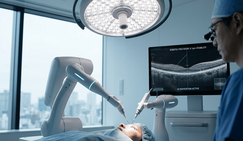
Advanced Diagnostics & Technology: Access to the latest high-resolution OCTs, visual field analyzers, and other cutting-edge equipment for precise IOP measurement and optic nerve assessment.
Expert Glaucoma Specialists: Highly trained and experienced ophthalmologists who are meticulous in their diagnostic approach and skilled in all forms of glaucoma management, from medication to advanced surgical techniques.
Uncompromising Safety & Quality Assurance: The Japanese medical system adheres to exceptionally high safety standards and rigorous quality control, ensuring reliable procedures and a secure medical environment.
The "Omotenashi" Experience: Patients benefit from Japan's unique spirit of selfless hospitality, translating into unparalleled attentiveness, respect, and personalized care throughout their medical journey.
Research & Innovation: Japan is actively involved in cutting-edge glaucoma research, including novel drug delivery systems and surgical approaches.
Proactive Steps for Your Eye Pressure and Glaucoma Risk
Understanding your eye pressure and its relationship to glaucoma is a vital step in preserving your vision. Regular, comprehensive eye examinations are essential, especially if you have risk factors for glaucoma (e.g., family history, age, high IOP).
If you are concerned about your eye pressure, have been diagnosed with ocular hypertension, or are seeking expert guidance on glaucoma management in Japan, we invite you to contact Ophthoagent. Our physician-led service provides expert guidance and connects you to leading glaucoma specialists and premier institutions in Japan, helping you navigate your path to preserving your vision.
This article was reviewed by
Dr. Daiki Sakai, MD